

- Cell Culture Media and Supplement
- Cell Dyes and Detection Assay Kits
- Matrices and Subctrates
- Cell Storage Media
- Instrument and Software
- Cell Isolation Products
- Density Gradient Media
- Laboratory Equipment
- Genome Editing and Molecular Tools
- Tissue and Cell Culture Dissociation Reagents
- Small Molecules
- Primary and Cultured Cells
- Culture and General Supplies

HOME >


제품특징

PneumaCult™ Airway Organoid kit는 인간 기관지 상피 세포 (HBEC)에서 유래한 기도 오가노이드의 효율적인 구축 및 분화를 위한 혈청 및 BPE가 없는 배양 배지 시스템입니다.
PneumaCult™ –Ex Plus Medium (ST05040)에서 2D 확장 후, PneumaCult™ Airway Organoid Seeding Medium을 사용하여 3D 오가노이드로의 배양을 시작한 다음 PneumaCult™ Airway Organoid Differentiation Medium으로 기도 오가노이드의 형태학적인 관련성을 갖추게 됩니다.
따라서 PneumaCult™ Airway Organoid kit는 기도 상피를 연구하기 위한 제품이며, 완전히 분화된 오가노이드는 ciliated cell 및 goblet cell같은 분화된 세포 유형으로 구성된 polarized airway epithelial cell layer로 둘러싸인 central lumen을 나타냅니다.
Advantages
- 인간 기도의 주요 특징을 재현한 3D 시스템
- PneumaCult™ –Ex Plus Medium (ST05040)와 호환되며, 인간 기도 상피 세포의 확장 및 분화를 위한 배지
- 일관된 결과 제공
- 편리한 형식과 사용하기 쉬운 프로토콜
General Workflow

Figure 1. Overview of the PneumaCult™ Culture System for Human Airway Organoid Generation
In the early two-dimensional expansion phase of the human airway organoid culture procedure, HBECs are expanded using PneumaCult™-Ex Plus Medium. The HBECs are then embedded into a Matrigel® dome and expanded for 4 - 7 days using PneumaCult™ Airway Organoid Seeding Medium. Following the expansion, the HBECs are differentiated using PneumaCult™ Airway Organoid Differentiation Medium for an additional 21+ days.
분화된 인간 기도 오가노이드

Figure 2. Fully Differentiated Human Airway Organoids Generated Using PneumaCult™ Airway Organoid Kit
(A) Bright-field image of airway organoids growing in PneumaCult™ Airway Organoid Seeding Medium at day 7 exhibit basal cell spheroid morphology. (B) Bright-field image of airway organoids differentiated in PneumaCult™ Airway Organoid Differentiation Medium at day 21 exhibit hollow lumens. (C) Airway organoid stained for ZO-1 (junction protein marker, red), MUC5AC (goblet cell marker, purple), AC-Tubulin (ciliated cell marker, green), and DAPI (nuclei, blue).

Figure 3. Forskolin-Induced Swelling of Airway Organoids
(A) Forskolin-treated organoids derived from healthy donors increased in size compared to the DMSO control, indicating functional CFTR protein expression. (B) Forskolin-induced swelling is lost in organoids derived from CF donors, but re-established in VX-809-treated airway organoids. Error bars represent ± 95% confidence interval for the mean (n=3). Bright-field images of airway organoids taken during the Forskolin swelling assay at (C) 0 hours and (D) 6 hours show organoid swelling after treatment.

Figure 4. Fully Differentiated Airway Organoids Retain Morphological Characteristics at Different Passages
The ciliated cell percentage in organoids grown from (A) healthy and (B) CF donors using PneumaCult™ Airway Organoid Kit increased from P3 to P5. The total and ciliated cells were counted using a hemocytometer. Error bars represent ± 95% confidence interval for the mean (n=3).
주문정보
| Cat No. | 품명 | 규격 | 제조사 | 제품정보 | 수량 | 견적문의 | |
|---|---|---|---|---|---|---|---|
| ST05060 | PneumaCult™ Airway Organoid Kit | 1 kit | StemCell Technologies | Serum- and BPE-free medium for efficient establishment and differentiation of airway organoids |   |
 카트담기 카트담기 견적문의 견적문의 |
| 전체선택 | |
|---|---|
|
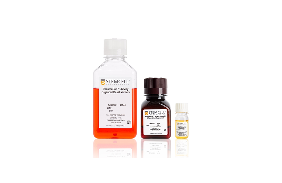
PneumaCult™ Airway Organoid Kit
|
관련제품
| Cat No. | 품명 | 규격 | 제조사 | 제품정보 | 수량 | 견적문의 | |
|---|---|---|---|---|---|---|---|
| ST07980 | Heparin Solution | 2 mL | StemCell Technologies | Cell culture supplement | |
카트담기 견적문의 |
|
| ST07925 | MesenCult™-ACF Chondrogenic Differentiation Kit | 100 mL | StemCell Technologies | Animal component-free medium for the differentiation of MSCs into chondrocytes | |
카트담기 견적문의 |
|
| ST05040 | PneumaCult™-Ex Plus | 1 kit | StemCell Technologies | Serum- and BPE-free medium for expansion of primary human bronchial and small airway epithelial cells (HBEC and HSAEC) | |
카트담기 견적문의 |
| 전체선택 | |
|---|---|
|
Heparin Solution
|
|
|
MesenCult™-ACF Chondrogenic Differentiation Kit
|
|
|
PneumaCult™-Ex Plus
|
최근 본 제품
제품 문의 02-3471-6500 042-824-7000
AS 문의 02-3471-8171 온라인 접수 