제품특징

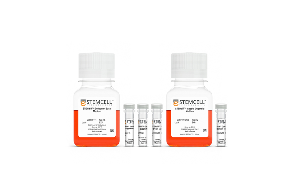
STEMdiff™ Gastric Organoid Differentiation Kit는 hPSC 계통 전반에 걸쳐 높은 효율성과 재현성으로 오가노이드의 성장 및 확장을 위한 media입니다.
Advantages
- 위의 상피(epithelium) 및 중간엽 (mesenchyme) 발달을 위한 시스템
- hES 및 iPSC의 효율적인 오가노이드 분화 가능
- 계대의 동결 보존이 가능해 실험적 유연성 확립 가능
- 무혈청 배지 시스템
General Workflow
- Initial condition (d-2): ~4000 hPSC aggregates/well in mTeSRTM1 or mTeSRTMPlus (50~200um)
- Previous to differentiation (d7): 85~90% confluence
- Gastric organoid (btw. d10~d26): matured gastric organoids surrounded by mesenchyme

Monolayer 에서 spheroid로 효율적 생성 가능


Figure 2. Efficient Formation and Budding of 3D Spheroids From 2D Posterior Foregut Monolayers
The formation and detachment of posterior foregut spheroids from differentiated 2D monolayers was observed between days 5 and 7 of differentiation using STEMdiff™ Gastric Organoid Kit. (A) Representative bright-field microscopy of day 7 spheroid morphologies presenting an outer polarized epithelium surrounding an inner cell mass. (B) Both ESC and iPSC-derived cultures demonstrate efficient spheroid formation upon posterior foregut formation. The total number of spheroids obtained per plate in a given differentiation is shown. (C) Immunofluorescence analysis of released posterior foregut spheroids after 7 days of differentiation. Spheroids express epithelial markers SOX2 and CADHERIN, and (D) a mesenchyme expressing vimentin, but not E-CADHERIN. Scale bars = 100 μm.
Gastric 오가노이드의 성숙 및 분화

Figure 3. Maturation and Differentiation of Human Gastric Organoids using STEMdiff™ Gastric Organoid Kit
Embedded PF spheroids shown in the image from day 10 were cultured in STEMdiff™ Gastric Organoid Medium and matured into gastric organoids surrounded by mesenchyme (day 20 - 26). Human gastric organoids were fully differentiated in STEMdiff™ Gastric Organoid Medium (day 26 - 34). Differentiated organoids showed a characteristic cystic morphology surrounded by mesenchymal cells. On day 34, formation of buds in the epithelium facing the lumen of the organoid was observed (white arrows). Scale bars = 500 μm.
Gastric 오가노이드의 빠른 확장성

Figure 4. Human Gastric Organoids Expand Rapidly in STEMdiff™ Gastric Expansion Organoid Medium
(A) Human gastric organoids derived from three cell lines were repeatedly passaged every 7 days in STEMdiff™ Gastric Organoid Expansion Medium. Representative images of human gastric organoids derived from WLS-1C iPS, H9 ES, and H1 ES cell lines at the end of passages 1, 3, 5, and 10 are shown. Matrigel® domes contained multiple organoids enriched in epithelial components with passaging progression, while losing the mesenchymal characteristic of early passages. Passage numbers are indicated at the top. Scale bars = 500 μm. (B) Human gastric organoids derived from multiple human ES (H1 & H9) and iPS (WLS-1C) cell lines undergo rapid expansion when maintained in STEMdiff™ Gastric Expansion Medium (n=1).
다양한 cell line에서 분화된 gastric organoid에서 gastric marker 확인 가능

Figure 5. Gene Expression Analysis of Human Gastric Organoids Cultured in Gastric Differentiation and Expansion Medium Shows that Organoids Express Gastric Markers
Gene expression analysis of human gastric organoids derived from four different cell lines (WLS-1C iPS, H9 ES, H1 ES, F022 iPS) cultured in either Differentiation Medium (left) for 34 days or Expansion Medium (right) for 0, 3, or 5 passages demonstrates comparable high expression of tight junction marker CLDN18, stem and progenitor cells markers LGR5, SOX2, and SOX9, and gland cell marker MUC6. High expression levels of MUC5AC and SST in differentiated and P0 organoids indicates the presence of pit and endocrine cells. Downregulation of these two markers was observed with extended culture and expansion to P3 and P5. High expression levels of GIF in both conditions indicates the presence of parietal cells that were not fully matured, as they lacked the expression of ATP4A. The presence of chief cell marker PGC was confirmed in differentiated and P0 organoids, and upregulation in organoids at P3 and P5 suggests enrichment of chief cells in expansion conditions. High expression levels of GASTRIN were observed in differentiated organoids and in human gastric organoids derived from WLS-1C and H9 lines in Expansion Medium; human gastric organoids derived from H1 and F022 show downregulation of GASTRIN in expansion conditions, suggesting a certain degree of inter-cell line variability in differentiating G cells. A commercially available mRNA sample of human adult stomach was used as a positive control. All samples were compared to undifferentiated hPSCs to calculate the relative gene expression (mean ± SD, n=1). hPSC line used and passage number (P) are given below the array.
Gastric-specific 마커의 발현의 면역조직화학적 확인 가능

Figure 6. Immunohistochemistry Confirms Expression of Gastric-Specific Markers in Human Gastric Organoids Cultured in Gastric Organoid Expansion Medium
Representative organoids in Expansion Medium at passage 5 expressed progenitor markers (A) SOX9, (B) SOX2, and (C) PDX1; (A,B&F) epithelial marker E-CADHERIN; (D) marker of proliferation Ki67; and (E) gastric tight junction marker CLDN18. (E) Presence of gland cells was detected by expression of MUC6 in the gland regions of the organoids. (F) Detection of scattered expression of PGC indicates differentiation of chief cells (n=2 - 5). Scale bars = 100 μm.


 카트담기
카트담기 견적문의
견적문의