제품특징
Advantages
- 다른 상업적 매체에 비해 더 많은 keratinocyte 획득 가능
- 환자 샘플에서 추출 효율성 향상
- 모든 계대에서 증식 세포의 동결 보존 가능
- Keratinocyte의 분화능 유지 가능
- 혈청 및 BPE 무함유 배양 배지
신생아 keratinocyte와 성인 keratinocyte 비교
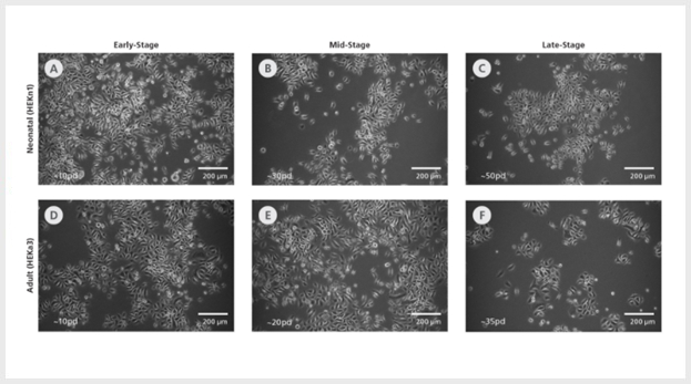
Figure 1. DermaCult™ Keratinocyte Expansion Medium Supports Long-Term Expansion of Primary Human Epidermal Keratinocytes
(A-C) Neonatal (HEKn1) and (D-E) adult (HEKa3) human epidermal keratinocytes (HEKs) cultured long-term in DermaCult™ Keratinocyte Expansion Medium maintain a cobblestone morphology throughout the culture period. The early-, mid-, and late-stages correspond to the number of population doublings reached. pd = population doublings. Scale bars = 200 μm. All images were taken using a 10X objective.
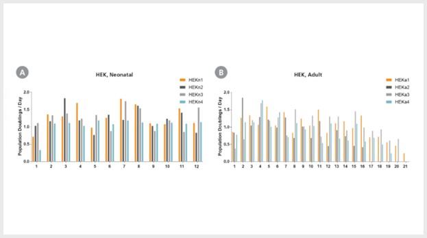
Figure 2. Primary Human Epidermal Keratinocytes Expanded in DermaCult™ Keratinocyte Expansion Medium Maintain High Growth Rates
(A) Neonatal and (B) adult HEKs cultured in DermaCult™ Keratinocyte Expansion Medium maintain a good proliferation rate over long-term culture, as observed by the population doublings /day at each passage. The neonatal cell lines were maintained until passage 12 while the adult cell lines were maintained until senescence.
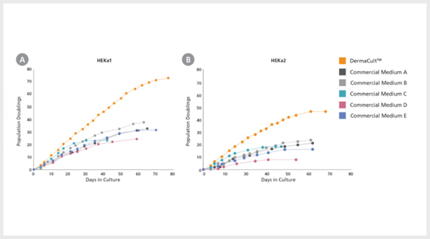
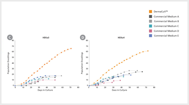
Figure 3. Primary Human Epidermal Keratinocytes Expanded in DermaCult™ Keratinocyte Expansion Medium Exhibit Increased Proliferation Rate Compared to Alternative Commercial Media
(A-D) Adult HEKs seeded and cultured in DermaCult™ Keratinocyte Expansion Medium show an increased proliferation rate and extended longevity compared to those cultured in 5 of the leading commercial media (n=4). Growth was assessed until senescence was reached.
DermaCultTM Keratinocyte Expansion Medium에서 확장된 세포는 조약돌 형태를 보이며 대표하는 주요 마커 (K14, p63, ITAG6 및 ITGB1)를 표현하며 air-liquid interface (ALI) 배양으로 분화에 적합합니다.
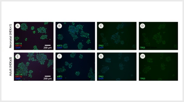
Figure 4. Primary Human Epidermal Keratinocytes Expanded in DermaCult™ Keratinocyte Expansion Medium Display Markers Characteristic of Basal Keratinocytes
(A-D) Neonatal and (E-H) adult HEKs maintained in DermaCult™ Keratinocyte Expansion Medium display characteristics of basal keratinocytes KRT14, KRT5, and TP63 (green) and absence of the suprabasal marker KRT10 (red), as observed through immunocytochemistry staining. Widespread co-labelling of the basal markers and the nuclear stain DAPI (blue) was observed. Scale bars = 200 μm. All images were taken using a 10X objective.
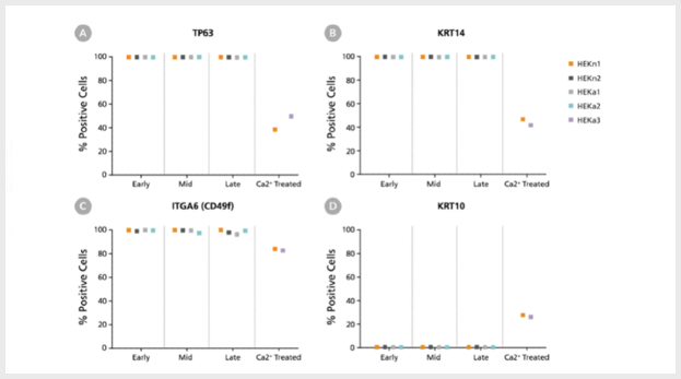
Figure 5. DermaCult™ Keratinocyte Expansion Medium Supports Expression of Key Protein Markers
Flow cytometry analysis of 2 neonatal (HEKn1, HEKn2) and 2 adult (HEKa1, HEKa2) HEK lines maintained in DermaCult™ Keratinocyte Expansion Medium was performed at early-, mid-, and late-stages. Ca2+ treated (differentiated) HEKs (HEKn1, HEKa3) were included as a control. A large percentage of the cells were positive for the basal markers (A) TP63, (B) KRT14, and (C) ITGA6 (CD49f) and negative for the differentiation marker (D) KRT10. In the Ca2+ treated HEKs, there is upregulation of KRT10 and downregulation of TP63, KRT14, and ITGA6.
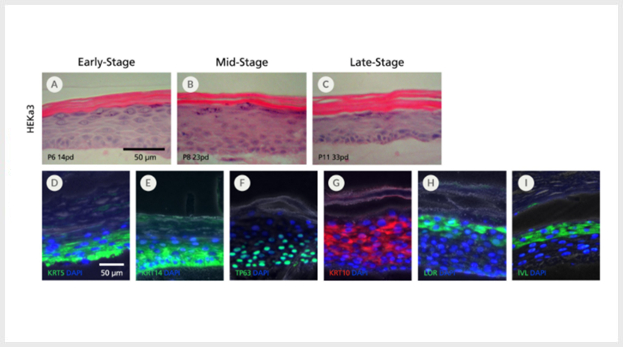
Figure 7. Human Epidermal Keratinocytes Maintained in DermaCult™ Keratinocyte Expansion Medium Differentiate at the Air-Liquid Interface
An adult keratinocyte line (HEKa3) cultured in DermaCult™ Keratinocyte Expansion Medium retains the ability to differentiate at the air-liquid interface (ALI) at (A) early-, (B) mid-, and (C) late-stages. H&E staining reveals a multi-layered structure with cuboidal basal cells and a clear cornified layer. The differentiated HEKs display protein markers characteristic of in vivo keratinocytes, including the expression of (D) KRT5, (E) KRT14, and (F) TP63 at the basal layers and (G) KRT10, (H) LOR, and (I) IVL at the suprabasal layers. Protein expression was visualized by immunohistochemistry. Fluorescent and phase contrast photos are overlaid. Scale bars = 50 μm. All images were taken using a 20X objective.







 카트담기
카트담기 견적문의
견적문의