


Cancer Research related Peptides
- 제품용도
- peptide를 이용한 다양한 실험에 활용 가능
- 브랜드
- ANASPEC
- 개요
- Cancer 연구를 위한 Purity: ≥95% by HPLC의 고순도 펩타이드

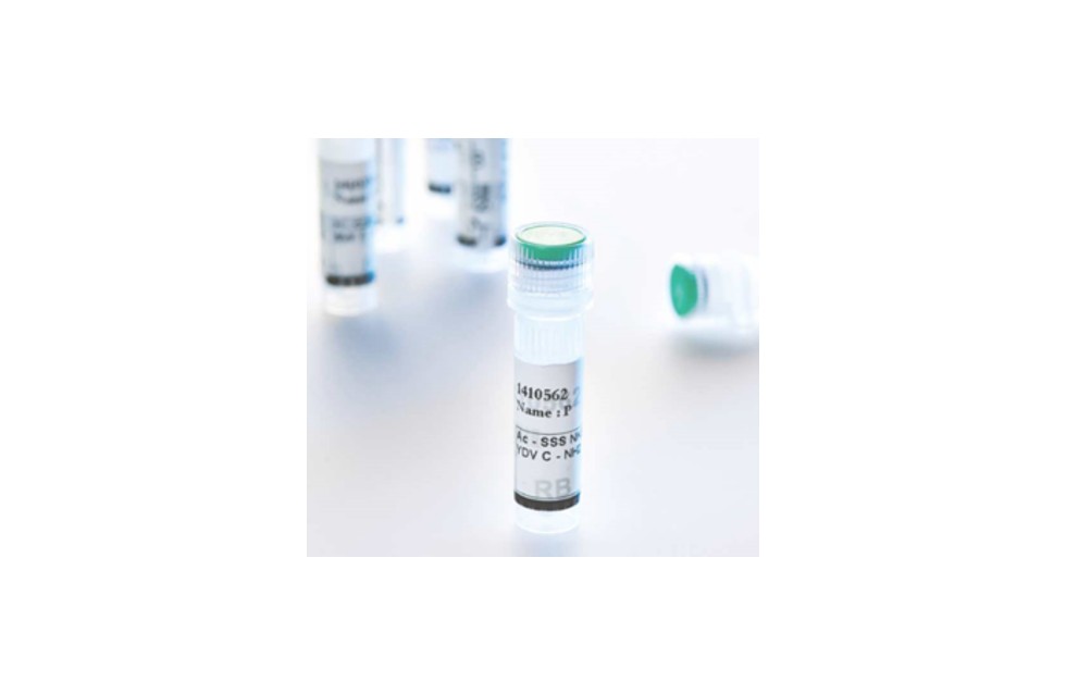

High Quality
- 고순도의 펩타이드 제공 (Purity: ≥95% by HPLC)
- ISO 9001 certified
- 경험과 지식이 풍부한 전문 인력에 의해 제조
맞춤형 합성 서비스 가능
| Cat No. | 품명 | 규격 | 제조사 | 제품정보 | 수량 | 견적문의 | |
|---|---|---|---|---|---|---|---|
| AS-61456 | [Asp370] - Tyrosinase (368 - 376) | 1 mg | Anaspec | This tumor antigenic peptide presented by HLA-A2 antigen to CTLs, is a tyrosinase fragment. Tyrosinase is a membrane-bound protein involved in the melanin synthesis pathway that is expressed by virtually all primary melanoma lesions and by most of metastatic lesions. |   |
 카트담기 카트담기 견적문의 견적문의 |
|
| AS-64798 | AH1 Sequence (6 - 14) | 1 mg | Anaspec | This AH1 sequence (amino acids 6 to 14) is the H2-Ld-restricted epitope derived from gp-70 (endogenous retroviral gene product envelope glycoprotein 70), which is expressed in CT26 (colon carcinoma cells) and numerous other tumor cell lines. | |
카트담기 견적문의 |
|
| AS-61616 | Bak BH3 | 1 mg | Anaspec | This peptide, derived from the BH3 domain of Bak (Flu-BakBH3), has been shown to have high-affinity binding to a surface pocket of the Bcl-XL protein that is essential for its death antagonist function. | |
카트담기 견적문의 |
|
| AS-64590 | Bak BH3 peptide, TAMRA - labeled | 1 mg | Anaspec | This peptide is derived from the BH3 domain of Bak (Flu-BakBH3). It has high-affinity binding to a surface pocket of the Bcl-XL protein that is essential for its death antagonist function. This peptide is labeled with 5-TAMRA on the N-terminus, Abs/Em= 541/568 | |
카트담기 견적문의 |
|
| AS-62266 | Bax BH3 peptide (55 - 74), wild type | 1 mg | Anaspec | This is a 20–amino acid Bax BH3 peptide (Bax 1) capable of inducing apoptosis in a variety of cell line models. In addition to disrupting Bax/Bcl-2 and Bax/Bcl-XL, it can promote cytochrome c release from isolated mitochondria. Bax BH3 belongs to the Bcl-2 protein family which consists of both pro-apoptotic and anti-apoptotic members. Bax BH3 acts to regulate apoptosis via governance of the 'intrinsic' pathway of cell death. | |
카트담기 견적문의 |
|
| AS-64082 | Bcl - 2 Binding Peptide, Bad BH3 Peptide | 1 mg | Anaspec | This is a bcl-2 binding peptide. This peptide is derived from the BH3 domain (a death domain) of Bad, amino acid residues 140 to 165. | |
카트담기 견적문의 |
|
| AS-61711 | Bid BH3 Peptide | 1 mg | Anaspec | BID is a pro-apoptotic member of the 'BH3-only' (BOPS) subset of the BCL-2 family of proteins that constitute a critical control point in apoptosis. Bid is the first of the BOPs reported to bind and activate Bcl-2, Bax, and Bak. Bid serves as a death-inducing ligand that moves from the cytosol to the mitochondrial membrane to inactivate Bcl-2 or to activate Bax. | |
카트담기 견적문의 |
|
| AS-61712 | Bid BH3, FAM labeled | 1 mg | Anaspec | This is a 5-FAM-labeled Bid BH3 peptide. Bid is a pro-apoptotic member of the 'BH3-only' subset of the BCL-2 family proteins that constitute a critical control point in apoptosis. Bid interconnects extrinsic pathway TNFR1 and Fas death signals to the mitochondrial amplification of the intrinsic pathway. The references listed below belong to the unlabeled Bid BH3. | |
카트담기 견적문의 |
|
| AS-62278 | Bim BH3, Peptide III | 1 mg | Anaspec | This Bim peptide belongs to the pro-apoptotic Bcl-2 family of proteins. | |
카트담기 견적문의 |
|
| AS-62279 | Bim BH3, Peptide IV | 1 mg | Anaspec | This Bim peptide belongs to the pro-apoptotic group of the Bcl-2 family of proteins. | |
카트담기 견적문의 |
|
| AS-24194 | c - Myc peptide epitope | 1 mg | Anaspec | c-Myc, the product of the c-myc proto-oncogene, is a helix-loop-helix leucine zipper phosphoprotein that regulates gene transcription in cell proliferation, cell differentiation and apoptosis. This peptide is a human c-myc epitope. | |
카트담기 견적문의 |
|
| AS-20590 | c - Myc peptide epitope | 5mg | Anaspec | c-Myc, the product of the c-myc proto-oncogene, is a helix-loop-helix leucine zipper phosphoprotein that regulates gene transcription in cell proliferation, cell differentiation and apoptosis. This peptide is a human c-myc epitope. | |
카트담기 견적문의 |
|
| AS-65302 | Enhanced Green Fluorescent Protein, EGFP (200 - 208) | 1 mg | Anaspec | This peptide is H2-Kd-restricted enhanced green fluorescent protein (EGFP)-derived peptide (200-208) and represents a CD8 T cell epitope. Immunization with this peptide stimulates IFNg production, thus making this peptide a model tumor antigen for the experimental development of antigen-specific vaccines against cancer. | |
카트담기 견적문의 |
|
| AS-61277 | G209 - 2M, gp100 (209 - 217) | 1 mg | Anaspec | This modified gp100 peptide amino acids 209 to 217 is a MHC-associated HLA-A2.1-restricted epitope derived from melanoma antigene. It can be processed, presented, and recognized by T cells. Alteration of the G209 peptide to G209-2M at the second amino acid changing threonine to a methionine was found to increase the affinity for MHC-associated HLA-A2.1 resulting in enhanced induction of T cells reactive to native gp100 | |
카트담기 견적문의 |
|
| AS-61515 | GAD65 (206 - 220) | 1 mg | Anaspec | A Glutamic acid decarboxylase 2 (GAD2) peptide corresponding to residues 206 to 220 of GAD65. GAD2 is presented to T cells in association with I-Ag7 MHC class II molecules. This peptide was also used as a part of Ig chimeras to test whether IL-10 interferes with expression of CTLA-4. | |
카트담기 견적문의 |
|
| AS-62589 | gp100 (25 - 33), human | 1 mg | Anaspec | This is amino acids 25 to 33 fragment of human melanoma antigen gp100. This H-2Db restricted epitope is recognized by T cells. The gp100-specific, H-2Db-restricted, CD8+ T cells are capable of recognizing B16 melanoma but not normal melanocytes. This peptide was used as an immunogen in multiple cancer immunotherapy studies. | |
카트담기 견적문의 |
|
| AS-61528 | HIF - 1 {alpha} (556 - 574) | 1 mg | Anaspec | This is a hypoxia-inducible factor-1 (HIF-1 a) 19-mer fragment. HIF-1 functions as master regulator of response to oxygen homeostasis. Hypoxia-induced gene expression is initiated when HIF-1 subunit is stabilized in response to a lack of oxygen. This part of HIF-1 binds to the von Hippel-Lindau factor (VHL) an E3 ubiquitin ligase, and the proline 564 is absolutely critical to the binding process | |
카트담기 견적문의 |
|
| AS-65581 | Human PD - L1 inhibitor I | 1 mg | Anaspec | Human PD-L1 inhibitor I is a peptide-based molecule. It binds to human PD-1 and inhibits PD-1/PD-L1 binding. This peptide, PDL-1 inhibitor I has anchor residues, WDY that influence binding of hPD-L1 to hPD-1. Developing inhibitors specifically blocking the PD-1/PD-L1 pathway has become a popular approach toward cancer treatment. | |
카트담기 견적문의 |
|
| AS-65582 | Human PD - L1 inhibitor II | 1 mg | Anaspec | Human PD-L1 inhibitor II is a peptide-based molecule. It binds to human PD-1 and inhibits PD-1/PD-L1 binding. This peptide, PDL-1 inhibitor I has anchor residues, WDY that influence binding of hPD-L1 to hPD-1. Developing inhibitors specifically blocking the PD-1/PD-L1 pathway has become a popular approach toward cancer treatment. | |
카트담기 견적문의 |
|
| AS-65583 | Human PD - L1 inhibitor III | 1 mg | Anaspec | Human PD-L1 inhibitor III is a peptide-based molecule. It binds to human PD-1 and inhibits PD-1/PD-L1 binding. This peptide, hPDL-1 inhibitor III has anchor residues (underlined), TEKDYRHGNIRMKLAYDL that influence binding of hPD-L1 to hPD-1. Developing inhibitors specifically blocking the PD-1/PD-L1 pathway has become a popular approach toward cancer treatment. | |
카트담기 견적문의 |
|
| AS-65584 | Human PD - L1 inhibitor IV | 1 mg | Anaspec | Human PD-L1 inhibitor IV is a peptide-based molecule. It binds to human PD-1 and inhibits PD-1/PD-L1 binding. This peptide, hPDL-1 inhibitor IV has anchor residues (underlined), GNWDYNSQRAQLYNQ that influence binding of hPD-L1 to hPD-1. Developing inhibitors specifically blocking the PD-1/PD-L1 pathway has become a popular approach toward cancer treatment. | |
카트담기 견적문의 |
|
| AS-64684 | Kisspeptin - 10 (Kp - 10), Metastin (110 - 119), amide, mouse, rat | 1 mg | Anaspec | This amidated peptide sequence is found in C-terminal residues 110 to 119 of the neurohormone Metastin (also referred to as Kisspeptin-10). This peptide increases plasma concentrations of GH (Growth Hormone) and LH (Luteinizing Hormone) in prepubertal dai | |
카트담기 견적문의 |
|
| AS-64240 | Kisspeptin - 10 (Kp - 10), Metastin (45 - 54) | 1 mg | Anaspec | This peptide sequence is found in residues 45 to 54 of Metastin (also referred to as Kisspeptin-10). This peptide increases plasma concentrations of GH (Growth Hormone) and LH (Luteinizing Hormone) in prepubertal dairy heifers. This peptide is the minimal sequence needed to activate GPR54 signaling, which may function in negatively regulating CXCR4’s role in programming tumor metastasis. Specifically, Kisspeptin-10 inhibits signaling and chemotaxis induced by SDF-1. | |
카트담기 견적문의 |
|
| AS-62169 | LyP - 1, Peptide 1 | 1 mg | Anaspec | LyP-1 recognizes lymphatics and tumor cells in certain tumors, but not lymphatics in normal tissues. Screening on breast carcinoma xenografts shows positive to cyclic 9-amino-acid peptide, LyP-1. The LyP-1 also recognizes an osteosarcoma xenograft, and spontaneous prostate and breast cancers in transgenic mice. LyP-1 peptide is detected in tumor structures that are positive for several lymphatic endothelial markers and negative for blood vessel markers. LyP-1 accumulates in the nuclei of the putative lymphatic cells, and in the nuclei of tumor cells. | |
카트담기 견적문의 |
|
| AS-61355 | MAGE - 3 (271 - 279) | 1 mg | Anaspec | This is a HLA-A*0201–restricted peptide derived from melanoma antigens encoded by MAGE-3. | |
카트담기 견적문의 |
|
| AS-61451 | MUC1, tandem repeat fragment | 1 mg | Anaspec | This sequence is the hallmark of MUC1 mucin. MUC1 is a highly glycosylated type I transmembrane glycoprotein with a unique extracellular domain consisting of a variable number of tandem repeats (VNTR) of this 20 amino acid peptide It is overexpressed on the cell surface of many human adenocarcinomas and hematological malignancies, including multiple myeloma and B-cell lymphoma, making MUC1 broadly applicable target for immunotherapeutic strategies | |
카트담기 견적문의 |
|
| AS-61334 | MUC5AC 3 | 1 mg | Anaspec | This glycopeptide is a 16-amino acid modified fragment of mucin 5/MUC5AC, where T* is a GalNac labeled threonine 3. Mucin-type linkages (GalNAc 1-O-Ser/Thr) are initiated by a family of glycosyltransferases known as the UDP-N-acetylgalactosamine:polypeptide N-acetylgalactosaminyltransferases (ppGaNTases). These enzymes transfer GalNAc from the sugar donor UDP-GalNAc to serine and threonine residues, forming an alpha anomeric linkage. The MUC5AC gene, which is mainly expressed in gastric and respiratory mucosae, exhibits 8 amino acid tandemly repeated domain, the consensus peptide TTSTTSAP. | |
카트담기 견적문의 |
|
| AS-61329 | MUC5AC, Analog 1 | 1 mg | Anaspec | This peptide is derived from the human mucin MUC5AC gene sequence. Data suggest that MUC5A and MUC5C are part of the same gene MUC5AC, which is distinct from MUC5B. The gene MUC5AC is mainly expressed in gastric, tracheo-bronchial mucosae and some tumors, it exhibits two kinds of deduced peptide domains, one of which is 8 amino acid tandemly repeated domain, a consensus peptide TTSTTSAP. | |
카트담기 견적문의 |
|
| AS-61333 | MUC5AC - 13 | 1 mg | Anaspec | This glycopeptide is an N-acetyl galactosamine (GalNAc)-modified MUC5AC mucin peptide containing the single site of threonine 13 labeled with GalNAc (T*). Polypeptide N-acetylgalactosaminyltransferase (ppGaNTase) catalyzes the transfer of GalNAc from the nucleotide sugar UDP-GalNAc to threonine. The MUC5AC gene is mainly expressed in gastric and tracheo-bronchial mucosae, and some tumors. | |
카트담기 견적문의 |
|
| AS-61332 | MUC5AC - 3/13 | 1 mg | Anaspec | This glycopeptide is a MUC5AC peptide with two sites (Thr3 and Thr13) labeled with GalNAc, where the asterisk denotes GalNAc-modified residues. Mucin type O-linked glycosylation is initiated by the action of a family of UDP-GalNAc: polypeptide N-acetylgalactosaminyltransferases (ppGaNTase), which catalyze the transfer of GalNAc from the nucleotide sugar UDP-GalNAc to the hydroxyl group of either serine or threonine. | |
카트담기 견적문의 |
|
| AS-64087 | Noxa A BH3 peptide, cell permeable | 1 mg | Anaspec | This cell permeable peptide is derived from the BH3 domain (a death domain) of Noxa A, amino acid residues 17 to 36. Eight D-Arginine residues and a Glycine linker residue are added to the amino terminal of the peptide. | |
카트담기 견적문의 |
|
| AS-62282 | Noxa BH3, Peptide 1 | 1 mg | Anaspec | This peptide belongs to the Bcl-2 family of proteins. Noxa gene encodes a Bcl-2 homology 3 (BH3)-only member of this family; it contains the BH3 region, not other BH domains. When ectopically expressed, Noxa undergoes BH3 motif-dependent localization to mitochondria, it interacts with anti-apoptotic Bcl-2 family members resulting in the activation of caspase-9. | |
카트담기 견적문의 |
|
| AS-65341 | Nuclear Factor (Erythroid - derived 2)like 2 (74 - 87); Nrf2 (74 - 87); NFE2L2 (74 - 87) | 5 mg | Anaspec | This peptide is derived from the Neh2 domain of nuclear factor (erythroid-derived 2)-like 2, or Nrf2. Nrf2 is a bZIP transcription factor that regulates the expression of antioxidative and cytoproptective genes. The DxETGE motif of this peptide binds Keap1 Kelch adaptor protein and displaces Nrf2 from Keap1. | |
카트담기 견적문의 |
|
| AS-62748 | Shepherdin (79 - 87) | 1 mg | Anaspec | This is amino acids 79 to 87 fragment of shepherdin, a novel peptidomimetic antagonist of the complex between Hsp90 and survivin, another key regulator of tumor cell viability. For its potent and broad antitumor activity, selectivity of action in tumor cells versus normal tissues, and inhibition of tumor growth in vivo without toxicity, shepherdin may offer a promising approach for rational cancer therapy. This sequence is also known as K79–K90. | |
카트담기 견적문의 |
|
| AS-65371 | Thrombospondin - derived Peptide; Cyclic CSVTCG | 1 mg | Anaspec | This peptide is derived from thrombospondin and represents a binding motif responsible for thrombospondin-CD36 interaction. It is cyclized through a disulfide bond. Thrombospondin is a matrix-bound glycoprotein involved in cancer metastasis, tumor adhesion, and angiogenesis. This peptide has been shown to competitively inhibit platelet aggregation and tumor metastasis. | |
카트담기 견적문의 |
|
| AS-61058 | TRP - 2 (180 - 188) | 1 mg | Anaspec | This peptide is derived from tyrosinase-related protein 2 (TRP2) residues 180-188. TRP2 belongs to the melanocyte differentiation antigens and has been implicated as a target for immunotherapy of human as well as murine melanoma. Studies show that this TRP2 derived peptide can bind to mouse and human MHC class I molecules. Immunization with TRP2 peptide loaded dendritic cells (DCs) results in effective induction of antitumor immunity. | |
카트담기 견적문의 |
|
| AS-64811 | TRP - 2, Tyrosinase - related Protein 2 (181 - 188) | 1 mg | Anaspec | This peptide is derived from tyrosinase-related protein 2 (TRP2) residues 181-188, and has been identified as the primary epitope of TRP2 recognized by anti-B16 melanoma cytotoxic T lymphocytes (CTLs). | |
카트담기 견적문의 |
|
| AS-60628 | VEGFR2/KDR Antagonist | 1 mg | Anaspec | This peptide is a specific VEGFR2/KDR heptapeptide antagonist, it binds VEGFR2 (KDR/flk), completely inhibiting VEGF binding to KDR and preventing VEGF-induced angiogenesis in-vivo. It specifically inhibits human endothelial cell proliferation in-vitro and totally abolishes VEGF-induced angiogenesis in-vivo. | |
카트담기 견적문의 |
|
| AS-62621 | WP9QY, TNF - alpha Antagonist | 1 mg | Anaspec | This cyclic peptide is designed to mimic the most critical tumor necrosis factor (TNF) recognition loop on TNF receptor I. It prevents interactions of TNF with its receptor. This TNF antagonist is a useful template for the development of small molecular inhibitors to prevent both inflammatory bone destruction and systemic bone loss in rheumatoid arthritis. | |
카트담기 견적문의 |
| 전체선택 | |
|---|---|
|
[Asp370] - Tyrosinase (368 - 376)
|
|
|
AH1 Sequence (6 - 14)
|
|
|
Bak BH3
|
|
|
Bak BH3 peptide, TAMRA - labeled
|
|
|
Bax BH3 peptide (55 - 74), wild type
|
|
|
Bcl - 2 Binding Peptide, Bad BH3 Peptide
|
|
|
Bid BH3 Peptide
|
|
|
Bid BH3, FAM labeled
|
|
|
Bim BH3, Peptide III
|
|
|
Bim BH3, Peptide IV
|
|
|
c - Myc peptide epitope
|
|
|
c - Myc peptide epitope
|
|
|
Enhanced Green Fluorescent Protein, EGFP (200 - 208)
|
|
|
G209 - 2M, gp100 (209 - 217)
|
|
|
GAD65 (206 - 220)
|
|
|
gp100 (25 - 33), human
|
|
|
HIF - 1 {alpha} (556 - 574)
|
|
|
Human PD - L1 inhibitor I
|
|
|
Human PD - L1 inhibitor II
|
|
|
Human PD - L1 inhibitor III
|
|
|
Human PD - L1 inhibitor IV
|
|
|
Kisspeptin - 10 (Kp - 10), Metastin (110 - 119), amide, mouse, rat
|
|
|
Kisspeptin - 10 (Kp - 10), Metastin (45 - 54)
|
|
|
LyP - 1, Peptide 1
|
|
|
MAGE - 3 (271 - 279)
|
|
|
MUC1, tandem repeat fragment
|
|
|
MUC5AC 3
|
|
|
MUC5AC, Analog 1
|
|
|
MUC5AC - 13
|
|
|
MUC5AC - 3/13
|
|
|
Noxa A BH3 peptide, cell permeable
|
|
|
Noxa BH3, Peptide 1
|
|
|
Nuclear Factor (Erythroid - derived 2)like 2 (74 - 87); Nrf2 (74 - 87); NFE2L2 (74 - 87)
|
|
|
Shepherdin (79 - 87)
|
|
|
Thrombospondin - derived Peptide; Cyclic CSVTCG
|
|
|
TRP - 2 (180 - 188)
|
|
|
TRP - 2, Tyrosinase - related Protein 2 (181 - 188)
|
|
|
VEGFR2/KDR Antagonist
|
|
|
WP9QY, TNF - alpha Antagonist
|
| Cat No. | 품명 | 규격 | 제조사 | 제품정보 | 수량 | 견적문의 | |
|---|---|---|---|---|---|---|---|
| ANA-AS-71120 | SensoLyte® AMC Caspase Profiling Kit *Fluorimetric* | 1 kit | Anaspec | The SensoLyte® Caspase Profiling Kit contains a series of AMC-based peptide substrates as fluorogenic indicators for assaying caspase protease activities. The kit contains a well-designed plate in which a series of AMC-based caspase substrates are coated with both positive and negative controls. It provides the best solution for profiling caspases or caspase inhibitors. *384-well plate format is available on custom basis. Kit Size: 2 plates | |
카트담기 견적문의 |
|
| ANA-AS-71118 | SensoLyte® Homogeneous AMC Caspase-3/7 Assay Kit | 1 kit | Anaspec | The SensoLyte® Homogeneous AMC Capase-3/7 Assay Kit uses Ac-DEVD-AMC as the fluorogenic indicator for assaying caspase-3/7 activities. Upon caspase-3/7 cleavage, Ac-DEVD-AMC generates the AMC fluorophore which has bright blue fluorescence and can be detected at Ex/Em=354 nm/442 nm. A bi-function assay buffer in this kit is designed to lyze the cells and measure the enzyme activity at the same time. Thus, this kit can measure caspase-3/7 activity in cell culture directly in a 96-well or 384-well plate without a time-consuming cell extraction step. In case the cells are cultured in larger plates or flasks, a lysis buffer and protocol for cell lysate preparation are also conveniently included in the kit. The kit is suitable for high throughput screening of apoptosis inducers and inhibitors. | |
카트담기 견적문의 |
|
| ANA-AS-71300 | Sensolyte® Cell Viability and Proliferation Assay Kit | 1 kit | Anaspec | The SensoLyte® Cell Viability and Proliferation Assay Kit provides researchers with a convenient one-solution and one-step assay to count living cells in a culture and continuously monitor cell proliferation over time by measuring cytoplasmic LDH activity. In this kit, resazurin is used as a sensitive fluorogenic indicator. Resazurin is converted to the strongly fluorescent resorufin (Ex/Em=560nm/590 nm) by cytoplasmic LDH. The kit can detect as few as 48 living cells with a linear range up to 5X10⁴ cells (R2=0.99). This kit can also be used for high throughput screening of cell proliferation or measuring cytotoxicity effect of a variety of compounds. 384-well or 1536-well format can be used with minor modifications. Kit Size: 2000 assays This assay is also available in a larger size (10,000 assays) | |
카트담기 견적문의 |
|
| ANA-AS-71302 | Sensolyte® Cell Cytotoxicity Assay Kit | 1 kit | Anaspec | The SensoLyte® Cell Cytotoxicity Assay Kit uses resazurin as a sensitive fluorogenic indicator (Ex/Em=560 nm/590 nm upon conversion) to measure LDH activity. The assay can be performed in a mixed population of damaged and viable cells, but it only measures the LDH released from damaged cells. The cytoplasmic LDH in living cells produces little signals under assay condition. There is no need for extra steps to separate living cells and supernatant. The fluorescent signal is proportional to the number of damaged cells (up to 2.5X10⁴ cell, r2>0.95) with the detection limit reaching 100 dead cells. The kit is suitable for high throughput screening of cytotoxicity of a variety of compounds. 384-well or 1536-well format can be used with minor modifications. Kit Size: 500 assays | |
카트담기 견적문의 |
| 전체선택 | |
|---|---|
|
SensoLyte® AMC Caspase Profiling Kit *Fluorimetric*
|
|
|
SensoLyte® Homogeneous AMC Caspase-3/7 Assay Kit
|
|
|
Sensolyte® Cell Viability and Proliferation Assay Kit
|
|
|
Sensolyte® Cell Cytotoxicity Assay Kit
|